CXCL10 prodotta dai follicoli piliferi induce l'infiltrazione di cellule Th1 e Tc1 nella fase acuta dell'alopecia areata, seguita da un prolungato accumulo di Tc1 nella fase cronica
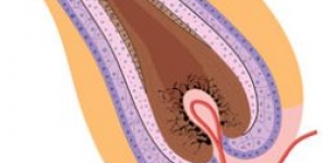
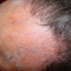
 L'alopecia areata (AA) è una malattia autoimmune, organo-specifica e cellulo-mediata. I linfociti T circondano densamente i bulbi piliferi lesionali, che vengono identificati istologicamente come "sciame d'api". Tuttavia, i meccanismi patogenetici dello "sciame d'api" sono tuttora incerti.
Obiettivo
Abbiamo studiato i meccanismi patologici dello "sciame d'api", concentrandosi sulla chemiotassi delle cellule T in modo tale che l'inibizione della chemiotassi possa essere fortemente candidata come nuovo trattamento per AA.
Metodi
Abbiamo studiato l'espressione dei recettori delle chemochine sulle cellule T ottenute dalle cellule mononucleate del sangue periferico (PBMCs) e dalle cellule infiltranti la pelle, di pazienti con AA. Inoltre, è stato anche eseguito il saggio di chemiotassi in tempo reale.
Risultati
Nelle PBMCs, la frequenza di cellule T CD4+ (Th1) CXCR3+ è stata significativamente superiore nel fase acuta di AA rispetto alla fase cronica di AA o al controllo sano, mentre le cellule T CD8+ (Tc1) CXCR3+ sono risultate significativamente aumentate nella fase cronica di AA. Nelle lesioni cutanee della fase acuta di AA, le cellule T CXCR3+CD4+ e CXCR3+CD8+ si sono infiltrate nella zona iuxta-follicolare. Nella fase cronica di AA, le cellule T CXCR3+CD8+ hanno dominato l'infiltrato intorno ai bulbi piliferi, contribuendo forse al prolungato stato di perdita dei capelli. I linfociti ottenuti dalla pelle lesionata in fase acuta di AA contenevano cellule T CXCR3+CD4+ e CXCR3+CD8+ in percentuali più elevate di quelle presenti nelle PBMCs, suggerendo un'emigrazione preferenziale dal sangue. Gli studi immunoistochimici e di real-time PCR hanno dimostrato che i follicoli piliferi nella fase acuta di AA esprimono un alto livello della chemochina CXCL10 associata a Th1. Con il saggio di chemiotassi, le PBMCs fresche e isolate da pazienti con fase acuta di AA hanno una maggiore velocità di chemiotassi verso CXCL10, con espressione aumentata di F-actina.
Conclusioni
Questi risultati suggeriscono che l'aumentata produzione di CXCL10 da parte di follicoli piliferi induce infiltrati preferenziali di cellule Th1 e Tc1 altamente chemoattratte nella fase acuta di AA, e inoltre l'infiltrazione di Tc1 rimane prolungata nella fase cronica.
Storia della pubblicazione:
Titolo: CXCL10 produced from hair follicles induces Th1 and Tc1 cell infiltration in the acute phase of alopecia areata followed by sustained Tc1 accumulation in the chronic phase
Rivista: Journal of Dermatological Science
Autori: Taisuke Ito, Hideo Hashizume, Takatoshi Shimauchi, Atsuko Funakoshi, Natsuho Ito, Hidekazu Fukamizu, Masahiro Takigawa, Yoshiki Tokura
Affiliazioni: Department of Dermatology, Hamamatsu University School of Medicine, 1-20-1 Handayama, Higashi-ku, Hamamatsu 431-1192, Japan
Department of Dermatology, Shimada Municipal Hospital, 1200-5 Noda, Shimada, Shizuoka 427-8502, Japan
Department of Reconstructive Plastic Surgery, Hamamatsu University School of Medicine, 1-20-1 Handayama, Higashi-ku, Hamamatsu 431-1192, Japan
Abstract:
Background
Alopecia areata (AA) is an organ-specific and cell-mediated autoimmune disease. T lymphocytes densely surround lesional hair bulbs, which is histologically referred to as "swarm of bees". However, pathomechanisms of "swarm of bees" are still uncertain.
Objective
We investigated the pathological mechanisms of "swarm of bees", focusing on T-cell chemotaxis so that inhibition of chemotaxis may be strong candidate of novel treatments for AA.
Methods
We investigate the expression of chemokine receptors on T cells obtained from peripheral blood mononuclear cells (PBMCs) and skin infiltrating cells in AA patients. In addition, real-time chemotaxis assay was also demonstrated.
Results
In PBMCs, the frequency of CXCR3+CD4+ T cells (Th1) was significantly higher in acute-phase AA than in chronic-phase AA or healthy control, while CXCR3+CD8+ T cells (Tc1) were significantly increased in chronic-phase AA. In the skin lesions of acute-phase AA, CXCR3+CD4+ and CXCR3+CD8+ T cells infiltrated in the juxta-follicular area. In chronic-phase AA, CXCR3+CD8+ T cells dominated the infiltrate around hair bulbs, possibly contributing to the prolonged state of hair loss. Lymphocytes obtained from a lesional skin of acute-phase AA contained CXCR3+CD4+ and CXCR3+CD8+ T cells at higher percentages than those of PBMCs, suggesting preferential emigration from the blood. Immunohistochemical and real-time RT-PCR studies demonstrated that hair follicles of acute-phase AA expressed a high level of Th1-associated chemokine CXCL10. By chemotaxis assay, freshly isolated PBMCs from acute-phase AA patients had a strong velocity of chemotaxis toward CXCL10 with increased expression of F-actin.
Conclusions
These results suggest that the increased production of CXCL10 from hair follicles induces preferential infiltrates of highly chemoattracted Th1 and Tc1 cells in the acute phase of AA, and Tc1 infiltration remains prolonged in the chronic phase.