Imaging ad alta risoluzione del carcinoma a cellule basali: un confronto fra microscopia multifotone e la microscopia confocale a riflettanza
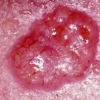
 Lo scopo di questo studio è stato quello di confrontare gli aspetti morfologici del carcinoma a cellule basali (BCC), valutandoli con due metodi di imaging diversi: la microscopia confocale a riflettanza in vivo (RCM) e la tomografia multifotone con l'applicazione dell'imaging di vita media di fluorescenza (MPT-FLIM).
Metodi
Lo studio ha riguardato 16 BCCs per i quali era disponibile una serie completa di immagini RCM e MPT-FLIM. È stata valutata la presenza di sette descrittori MPT-FLIM. La presenza di sette parametri equivalenti RCM è stata ottenuta in accordo con la loro estensione. Il test del Chi-quadro con il test esatto di Fisher e il coefficiente di correlazione dei ranghi di Spearman sono stati determinati tra i punteggi MPT-FLIM e quelli RCM rettificati.
Risultati
I descrittori MPT-FLIM e RCM di BCC sono stati accoppiati per far corrispondere i descrittori che definiscono le stesse strutture patologiche. Il confronto ha incluso: il flusso e allineamento delle cellule allungate, il flusso con direzioni multiple e con doppio allineamento, palizzamento (RCM) e palizzamento (MPT-FLIM), Tipiche isole tumorali, e isole di cellule circondate da fibre, sagome scure e isole fantasma, cellule luminose paffute e melanofagi, vasi (RCM), e vasi (MPT-FLIM). I parametri risultati significativamente correlati sono stati i Melanofagi/cellule luminose paffute, Doppio allineamento/Flusso con direzioni multiple, e Palizzamento (MPT-FLIM)/Palizzamento (RCM).
Conclusione
Secondo i nostri dati, entrambi i metodi sono adatti per raffigurare le caratteristiche dei BCCs. La concordanza tra MPT-FLIM e RCM è alta, con alcune limitazioni dovute alle differenti tecniche tra i due dispositivi. La difficoltà più alta, quando si confrontano le immagini generate dalle due modalità di imaging, è rappresentata dal loro diverso campo di vista.
Storia della pubblicazione:
Titolo: High-resolution imaging of basal cell carcinoma: a comparison between multiphoton microscopy with fluorescence lifetime imaging and reflectance confocal microscopy
Rivista: Skin Research and Technology. doi: 10.1111/j.1600-0846.2012.00661.x
Autori: Marco Manfredini, Federica Arginelli, Christopher Dunsby, Paul French, Clifford Talbot, Karsten König, Giovanni Pellacani, Giovanni Ponti, Stefania Seidenari
Affiliazioni: Department of Dermatology, University of Modena and Reggio Emilia, Modena, Italy
Department of Physics, Imperial College London, London, UK
Department of Biophotonics and Lasertechnology, Saarland University, Saarbrücken, Germany
JenLab GmbH, Jena, Germany
Department of Clinical and Diagnostic Medicine and Public Health, University Hospital of Modena and Reggio Emilia, Italy
Abstract:
Aims
The aim of this study was to compare morphological aspects of basal cell carcinoma (BCC) as assessed by two different imaging methods: in vivo reflectance confocal microscopy (RCM) and multiphoton tomography with fluorescence lifetime imaging implementation (MPT-FLIM).
Methods
The study comprised 16 BCCs for which a complete set of RCM and MPT-FLIM images were available. The presence of seven MPT-FLIM descriptors was evaluated. The presence of seven RCM equivalent parameters was scored in accordance to their extension. Chi-squared test with Fisher's exact test and Spearman's rank correlation coefficient were determined between MPT-FLIM scores and adjusted-RCM scores.
Results
MPT-FLIM and RCM descriptors of BCC were coupled to match the descriptors that define the same pathological structures. The comparison included: Streaming and Aligned elongated cells, Streaming with multiple directions and Double alignment, Palisading (RCM) and Palisading (MPT-FLIM), Typical tumor islands, and Cell islands surrounded by fibers, Dark silhouettes and Phantom islands, Plump bright cells and Melanophages, Vessels (RCM), and Vessels (MPT-FLIM). The parameters that were significantly correlated were Melanophages/Plump Bright Cells, Aligned elongated cells/Streaming, Double alignment/Streaming with multiple directions, and Palisading (MPT-FLIM)/Palisading (RCM).
Conclusion
According to our data, both methods are suitable to image BCC's features. The concordance between MPT-FLIM and RCM is high, with some limitations due to the technical differences between the two devices. The hardest difficulty when comparing the images generated by the two imaging modalities is represented by their different field of view.
https://www.youtube.com/@djfdm