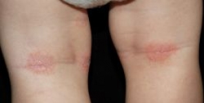
Modelli di distribuzione PGP 9.5 in biopsie di lesioni precoci della dermatite atopica
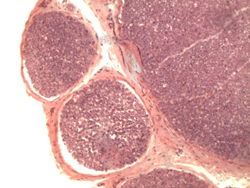 Le fibre nervose periferiche sono spesso aumentate nella cute lesionata di pazienti con dermatite atopica (AD). Abbiamo cercato di studiare i profili delle fibre nervose, usando PGP 9.5 come marker neuronale, in lesioni precoci da AD in 10 pazienti, rispetto alla pelle non lesionata degli stessi pazienti e alla pelle da controlli sani.
Il numero di profili di fibre nervose positivi al PGP 9.5 non è stato differente nelle biopsie effettuate da pelle con AD apparentemente normale e da controlli sani. Il numero totale di profili di fibre nervose positivi al PGP 9.5 nelle sezioni di pelle intera è stato maggiore sia nell'epidermide che nel derma del gruppo di biopsie cutanee effettuate da lesioni precoci di pazienti con AD. Inoltre, il numero di cellule dendritiche epidermiche positive al PGP 9.5 è stato superiore nella pelle con AD.
Sembra ragionevole che le fibre nervose positive al PGP 9.5 e le cellule dendritiche positive al PGP 9.5 abbiano ruoli patologici nell'AD. I risultati potrebbero servire come base per ulteriori studi nella valutazione di nuovi approcci diagnostici e terapeutici.
Fonte:
Titolo: PGP 9.5 distribution patterns in biopsies from early lesions of atopic dermatitis
Rivista: Archives of Dermatological Research. doi: 10.1007/s00403-012-1246-0
Autori: Lennart Emtestam, Lena Hagströmer, Ying-Chun Dou, Karin Sartorius, Olle Johansson
Affiliazioni: Section of Dermatology I 43, Department of Medicine, Karolinska Institutet, Huddinge University Hospital, 141 86 Stockholm, Sweden
Section of Dermatology, Department of Clinical Science and Education, Karolinska Institutet, Södersjukhuset, Stockholm, Sweden
The Experimental Dermatology Unit, Department of Neuroscience, Karolinska Institutet, Stockholm, Sweden
Abstract:
Peripheral nerve fibres are often increased in lesional skin of atopic dermatitis (AD) patients. We attempted to study nerve fibre profiles, using PGP 9.5 as neuronal marker, in early AD lesions in 10 patients, as compared to non-lesional skin in the same patients and skin from healthy controls. The number of PGP 9.5-positive nerve fibre profiles was not different in the biopsies taken from normal-looking AD skin and healthy controls. The total number of PGP 9.5-positive nerve fibre profiles in the whole skin sections was higher in both the epidermis and the dermis in the group of skin biopsies taken from early lesions of AD patients. Further, the number of epidermal PGP 9.5-positive dendritic cells was increased in AD skin. It seems reasonable that PGP 9.5-positive nerve fibres and PGP 9.5-positive dendritic cells have pathological roles in AD. The findings might serve as a basis for further studies in evaluating novel diagnostic and therapeutic approaches.
