Identificazione e suscettibilità antifungina di funghi isolati da dermatomicosi
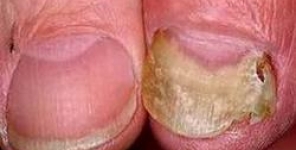
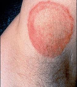 Le dermatomicosi sono infezioni fungine superficiali della pelle, dei capelli e delle unghie che colpiscono più del 20-25% delle persone in tutto il mondo. Queste infezioni possono essere causate da dermatofiti, lieviti e funghi filamentosi non dermatofiti (NDFF) e sono considerate un problema di salute pubblica. Nonostante questo, solo pochi studi hanno esaminato la prevalenza e la suscettibilità antifungina degli agenti causali di dermatomicosi, nei paesi in via di sviluppo.
Obiettivi
Gli obiettivi di questo studio sono stati quelli di identificare e determinare il profilo di suscettibilità antimicotico dei lieviti e dei funghi filamentosi isolati dalle dermatomicosi in Uberaba, Minas Gerais e in Brasile.
Metodi
I campioni sono stati ottenuti da pazienti affetti da dermatomicosi diagnosticate clinicamente e confermate in laboratorio, tra il luglio 2009 e il luglio 2011. L'identificazione fungina si è basata su metodi classici mentre il test di suscettibilità antifungina è stato eseguito con il metodo di microdiluizione in brodo.
Risultati
Dei 216 isolati fungini, 116 (53.8%) erano lieviti, 70 (32.4%) dermatofiti e 30 (13.8%) NDFF. L'onicomicosi è stata la condizione clinica più comune. La Candida parapsilosis (24.1%) e il Trichophyton rubrum (17.1%) sono stati i funghi più frequentemente isolati. Il voriconazolo, il ketoconazolo e l'itraconazolo sono risultati gli agenti antifungini più potenti contro i lieviti, mentre la terbinafina, l'itraconazolo e il voriconazolo hanno avuto un'alta attività in vitro nei confronti dei dermatofiti. Nel complesso, gli agenti antifungini hanno avuto poca o nessuna attività contro i NDFF e le massime concentrazioni minime inibenti sono state quelle nei confronti di Fusarium spp.
Conclusione
I lieviti, in particolare C. parapsilosis, svolgono un ruolo importante come agenti causali delle dermatomicosi, nella nostra regione. I nostri risultati suggeriscono che il test di sensibilità antimicotica accoppiato con una corretta identificazione dei funghi può essere utile per aiutare i medici nel determinare la terapia appropriata nei confronti delle dermatomicosi.
Storia della pubblicazione:
Titolo: Identification and antifungal susceptibility of fungi isolated from dermatomycoses
Rivista: Journal of the European Academy of Dermatology and Venereology. doi: 10.1111/jdv.12151
Autori: L.B. Silva, D.B.C. de Oliveira, B.V. da Silva, R.A. de Souza, P.R. da Silva, K. Ferreira-Paim, L.E. Andrade- Silva, M.L. Silva-Vergara, A.A. Andrade
Affiliazioni:Laboratory of Microbiology, Institute of Biological and Natural Sciences, Federal University of Triângulo Mineiro, Uberaba, Minas Gerais, Brazil Infectious Diseases Unit, Federal University of Triângulo Mineiro, Uberaba, Minas Gerais, Brazil
Abstract:
Background Dermatomycoses are superficial fungal infections of the skin, hair and nails that affect more than 20–25% of the people worldwide. These infections can be caused by yeasts, dermatophytes and non- dermatophyte filamentous fungi (NDFF) and are considered a public health problem. Despite this, few studies have investigated the prevalence and antifungal susceptibility of causative agents of dermatomycoses in the developing world. Objectives The aims of this study were to identify and determine the antifungal susceptibility profile of yeast and filamentous fungi isolated from dermatomycoses in Uberaba, Minas Gerais, Brazil. Methods Specimens were obtained from patients with clinically diagnosed and laboratory confirmed dermatomycosis between July 2009 and July 2011. Fungal identification was based on classical methods and antifungal susceptibility testing was performed by broth microdilution method. Results Of the 216 fungal isolates, 116 (53.8%) were yeasts, 70 (32.4%) dermatophytes and 30 (13.8%) NDFF. Onychomycosis was the most common clinical condition. Candida parapsilosis (24.1%) and Trichophyton rubrum (17.1%) were the fungi most frequently isolated. Voriconazole, ketoconazole and itraconazole were the most potent antifungal agents against yeast, whereas terbinafine, voriconazole and itraconazole had a high in vitro activity against dermatophytes. Overall, the antifungal agents had little or no activity against NDFF and the highest minimum inhibitory concentrations were those against Fusarium spp. Conclusion Yeasts, particularly C. parapsilosis, play an important role as causative agents of dermatomycosis in our region. Our results suggest that the antifungal susceptibility testing coupled with proper identification of the fungi may be useful to assist clinicians in determining the appropriate therapy for dermatomycoses.
