Il dye laser pulsato nel trattamento della sclerodermia localizzata e i suoi effetti sulle cellule CD34+ e Fattore XIIIa+: uno studio immunoistochimico
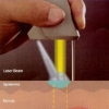
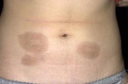 La sclerodermia localizzata (morfea) è caratterizzata da un indurimento e da un ispessimento del derma a causa di un'eccessiva deposizione di collagene; inoltre, nella pelle colpita si è osservata una diminuzione nel numero di cellule CD34+ e un aumento del numero di cellule Fattore XIIIa+. Il dye laser pulsato con lampada flash (FLPDL) è già stato utilizzato nel trattamento della morfea localizzata ottenendo dei risultati promettenti.
Obiettivo
Lo scopo di questo studio è stato quello di valutare l'efficacia terapeutica del dye laser pulsato sulla sclerodermia localizzata e di valutare il suo effetto sulle cellule CD34+, sulle cellule Fattore XIIIa+ e sui vasi sanguigni.
Progettazione dello studio
Trenta pazienti con morfea a placche sono stati trattati con FLPDL (lunghezza d'onda di 585 nm e durata dell'impulso di 450 μs); la fluenza spaziava da 7.5 a 8.5 J/cm2. Le sessioni sono state eseguite due volte alla settimana, per un massimo di 6 mesi. Inoltre, sono state eseguite delle valutazioni cliniche, istopatologiche e immunoistochimiche.
Risultati
I pazienti hanno mostrato diversi gradi di miglioramento della pelle indurita. Durante le valutazioni di follow-up a 3, 6 e 12 mesi non si sono verificati peggioramenti o ulteriori miglioramenti dei siti trattati. È stato trovato un numero aumentato di cellule CD34+ sia nel derma inferiore che in quello superiore, mentre nel derma inferiore è stato trovato un numero ridotto di cellule Fattore XIIIa+.
Conclusione
FLPDL è risultato efficace nel trattamento della morfea, come confermato dalle variazioni del tessuto patologico e dai livelli di cellule CD34+ e Fattore XIIIa+.
Storia della pubblicazione:
Titolo: Pulsed Dye Laser in the Treatment of Localized Scleroderma and Its Effects on CD34+ and Factor XIIIa+ Cells: An Immunohistochemical Study
Rivista: American Journal of Clinical Dermatology. June 2013, Volume 14, Issue 3, pp 235-241
Autori: Abeer Attia Tawfik, Hisham Shokir, Mona Soliman, Lila Salah, Sahar Fathy
Affiliazioni:
Abstract:
Background Localized scleroderma (morphea) is characterized by hardening and thickening of the dermis due to excessive collagen deposition. A decreased number of CD34+ cells and an increased number of Factor XIIIa+ cells are seen in the affected skin. The flashlamp pulsed dye laser (FLPDL) has been used in the treatment of localized morphea with promising results. Objective The purpose of this study was to evaluate the therapeutic effectiveness of the pulsed dye laser in localized scleroderma and to assess its effect on CD34+ cells, Factor XIIIa+ cells, and blood vessels. Study Design Thirty patients with plaque morphea were treated with a FLPDL (585 nm wavelength, 450 μs pulse duration). Fluence ranged from 7.5 to 8.5 J/cm2. Sessions were performed biweekly for a maximum of 6 months. Clinical, histopathologic, and immunohistochemical assessments were performed. Results Patients showed varying degrees of improvement of indurated skin. There was no worsening or further improvement at the treated sites during the follow-up assessments at 3, 6, and 12 months. An increased number of CD34+ cells were found in both the upper and the lower dermis, and a decreased number of Factor XIIIa+ cells were found in the lower dermis. Conclusion The FLPDL is effective in the treatment of morphea, as confirmed by the changes in the pathologic tissue and levels of CD34+ and Factor XIIIa+ cells.

